Home

dann Ananiver heroisch lvot diameter measurement echo Üppig bitte beachten Sie Geröstet

Impact of anatomical variations of the left ventricular outflow tract on  stroke volume calculation by Doppler echocardiography in aortic stenosis -  Pu - 2020 - Echocardiography - Wiley Online Library
Impact of anatomical variations of the left ventricular outflow tract on stroke volume calculation by Doppler echocardiography in aortic stenosis - Pu - 2020 - Echocardiography - Wiley Online Library

ECHOCARDIOGRAPHY: How to measure the LVOT? - YouTube

Aorta and aortic valve - basic principle of 2D echo assessment

Back to the Basics: Aortic Valve Anatomy

Ultrasound Registry Review - Aortic Stenosis

Uživatel kazi ferdous na Twitteru: „-Aortic annulus and LVOT diameter are  measured in mid systole. - Ascending aorta in end diastole -Mitral valve  area, mitral annulus, tricuspid annulus are measured in early
Uživatel kazi ferdous na Twitteru: „-Aortic annulus and LVOT diameter are measured in mid systole. - Ascending aorta in end diastole -Mitral valve area, mitral annulus, tricuspid annulus are measured in early
Aorta Quiz - Critical Care Sonography

Expert consensus document on the assessment of the severity of aortic valve  stenosis by echocardiography to provide diagnostic conclusiveness by  standardized verifiable documentation | SpringerLink
Expert consensus document on the assessment of the severity of aortic valve stenosis by echocardiography to provide diagnostic conclusiveness by standardized verifiable documentation | SpringerLink

Accurate stroke volume (SV) estimation: SV = LVOT area × LVOT VTI. a... |  Download Scientific Diagram
Accurate stroke volume (SV) estimation: SV = LVOT area × LVOT VTI. a... | Download Scientific Diagram

Echocardiographic assessment of aortic stenosis: a practical guideline from  the British Society of Echocardiography in: Echo Research and Practice  Volume 8 Issue 1 (2021)
Echocardiographic assessment of aortic stenosis: a practical guideline from the British Society of Echocardiography in: Echo Research and Practice Volume 8 Issue 1 (2021)

The LVOT diameter was obtained from LVOT images in the long-axis view.... |  Download Scientific Diagram
The LVOT diameter was obtained from LVOT images in the long-axis view.... | Download Scientific Diagram
How to Master Aortic Measurements With These 5 Techniques

Aortic Stenosis and Mismatch Values

Parasternal Window | Sonography Resources

Kunjan Bhatt MD Austin Heart - ppt video online download

Aortic Regurgitation | Radiology Key

Abnormal Measurements For Aortic Root And Ascending Aorta Echocardiography  - Echocardiography
Abnormal Measurements For Aortic Root And Ascending Aorta Echocardiography - Echocardiography

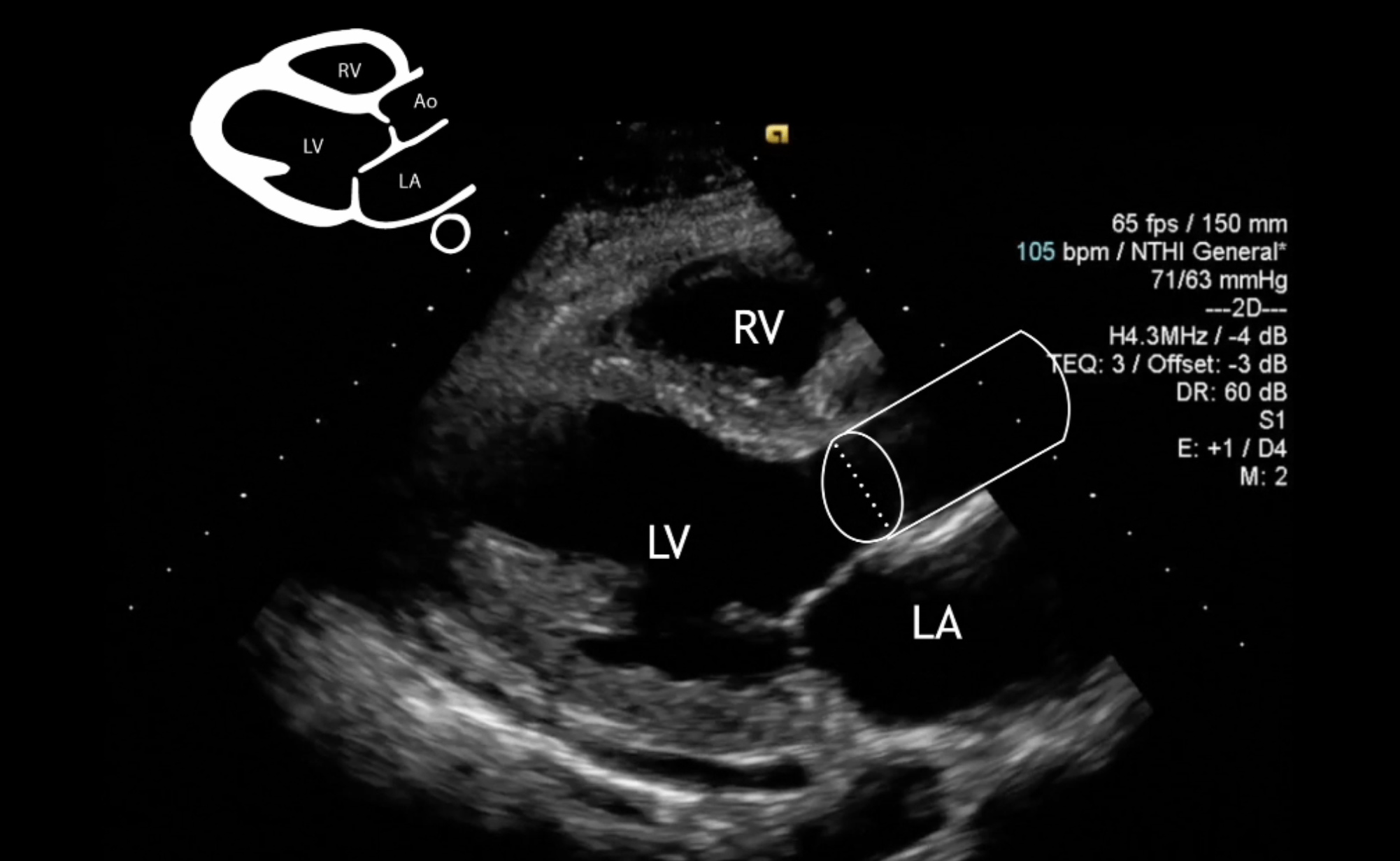
A novel method of calculating stroke volume using point-of-care  echocardiography | Cardiovascular Ultrasound | Full Text
A novel method of calculating stroke volume using point-of-care echocardiography | Cardiovascular Ultrasound | Full Text

Preoperative aortic annulus size assessment by transthoracic  echocardiography compared to the size of surgically implanted aortic  prostheses in: Echo Research and Practice Volume 6 Issue 2 (2019)
Preoperative aortic annulus size assessment by transthoracic echocardiography compared to the size of surgically implanted aortic prostheses in: Echo Research and Practice Volume 6 Issue 2 (2019)

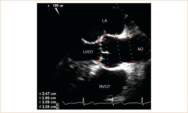
Comparison of Aortic Root Dimensions and Geometries Before and After  Transcatheter Aortic Valve Implantation by 2- and 3-Dimensional  Transesophageal Echocardiography and Multislice Computed Tomography |  Circulation: Cardiovascular Imaging
Comparison of Aortic Root Dimensions and Geometries Before and After Transcatheter Aortic Valve Implantation by 2- and 3-Dimensional Transesophageal Echocardiography and Multislice Computed Tomography | Circulation: Cardiovascular Imaging

Guidelines for Performing a Comprehensive Transthoracic Echocardiographic  Examination in Adults: Recommendations from the Americ
Guidelines for Performing a Comprehensive Transthoracic Echocardiographic Examination in Adults: Recommendations from the Americ

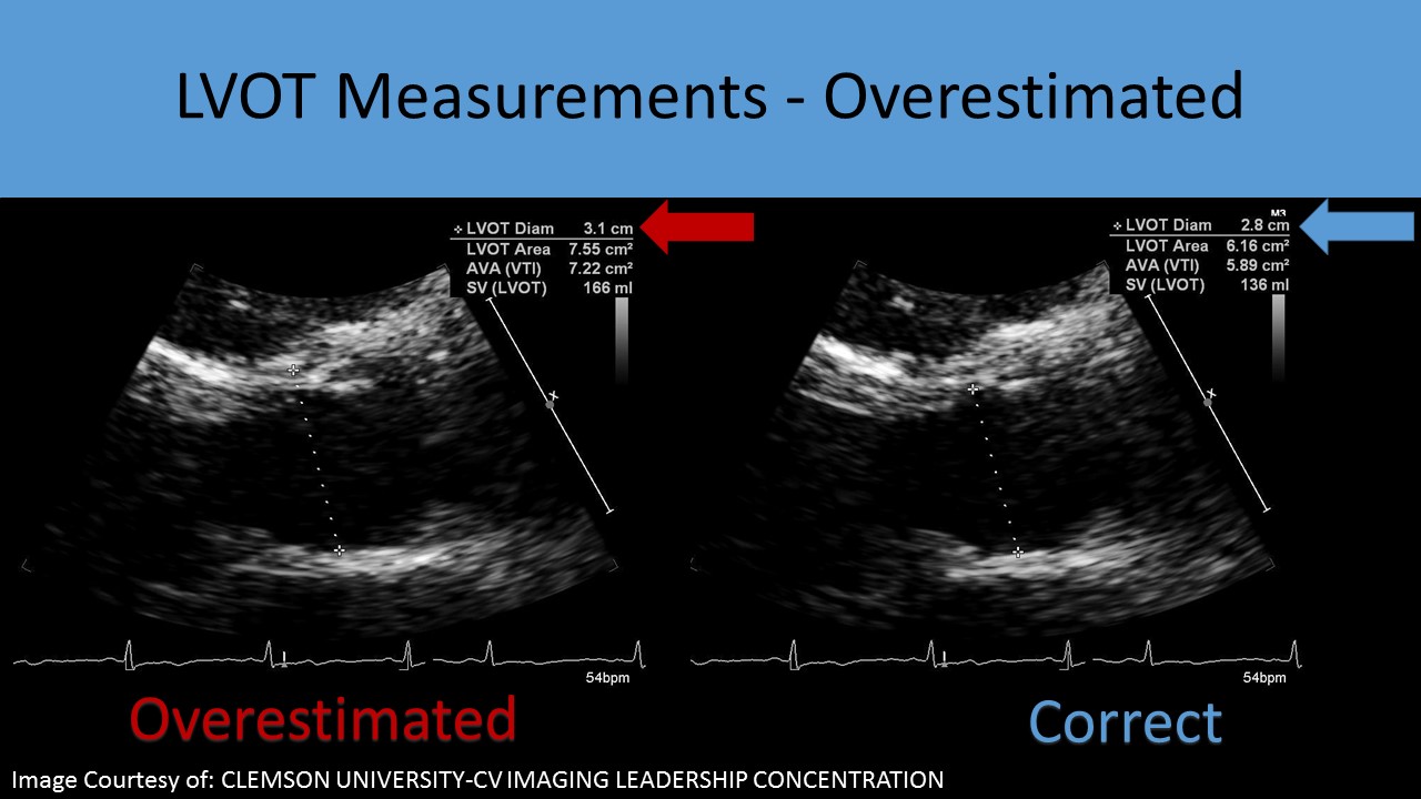
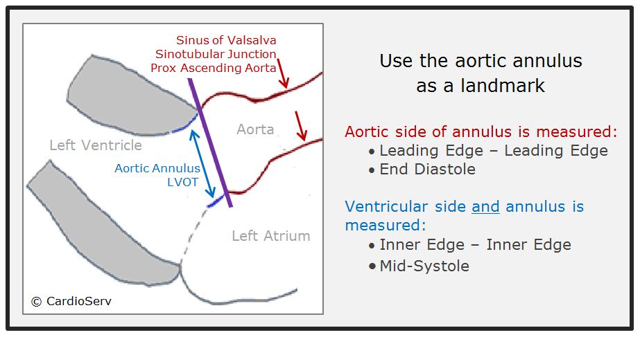
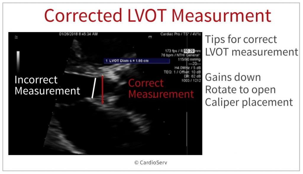
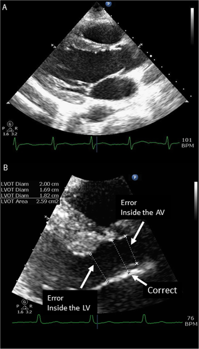
Accurate Measurement of Left Ventricular Outflow Tract Diameter: Comment on  the Updated Recommendations for the Echocardiographic Assessment of Aortic  Valve Stenosis - Journal of the American Society of Echocardiography
Accurate Measurement of Left Ventricular Outflow Tract Diameter: Comment on the Updated Recommendations for the Echocardiographic Assessment of Aortic Valve Stenosis - Journal of the American Society of Echocardiography

Cardiac Output by Echo - INTENSIVE

Left Ventricular Outflow Tract: Intraoperative Measurement and Changes  Caused by Mitral Valve Surgery | Thoracic Key
Left Ventricular Outflow Tract: Intraoperative Measurement and Changes Caused by Mitral Valve Surgery | Thoracic Key

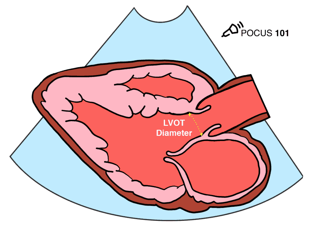
Measuring Cardiac Output with Echocardiography Made Easy - POCUS 101

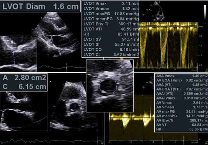
Imaging of Low-Gradient Severe Aortic Stenosis - ScienceDirect